Patients with epilepsy have ‘spontaneous abnormal synchronized hyperexcitation’ in the brain – so called seizures. Seizures can occur any time during the day or night. For a patient, not knowing when he or she may suddenly lose consciousness or control over his or her body has a significant impact on daily life. About 0.7% of the population has epilepsy and of these more than 30% are resistant to any current medication.
Epileptic seizures may arise from almost anything that damages the brain; brain trauma, stroke, infections and tumors as well as inherited genetic mutations. Epileptic seizures per se may also lead to brain damage.
The inflammatory reaction
For some time now, the inflammatory reaction in the brain following epileptic seizures has gained increased interest.
For some time now, the inflammatory reaction in the brain following epileptic seizures has gained increased interest. Modulations of several inflammatory signaling pathways have succeeded in reducing brain inflammation.
However, since we know now that the immune response in the brain includes both damaging and restorative mechanisms, there is a need for improvement and development of more selective immune modulating therapies.
When evaluating one of our anti-inflammatory therapies in rats with temporal lobe seizures, we came to discuss our project with Dr Ulrica Englund-Johanson at the Dept of Ophthalmology in Lund and realized that a possible immune response in the retina following epileptic seizures had never been evaluated.
The retina is a remote extension of the brain and an intracranial spread of the inflammation to the retina may therefore be possible. Since we also know that epileptic seizures lead to an acute immune reaction in the blood, the inflammation may also spread systemically to other organs including the eyes.
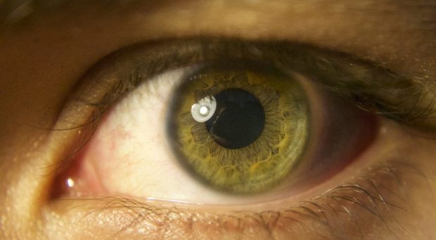
The eyes and epilepsy
The study that we designed was explorative, since we had very little clinical evidence to base our assumptions on. Patients with epilepsy seldom complain about visual problems, except for those with seizures originating from the vision-regulated occipital lobe.
There are several studies showing visual disturbances and structural changes of the different layers in the retina in patients with epilepsy due to anti-epileptic medications.
There are, to our knowledge, no publications that have described damage in retinal specimens from patients with epilepsy. However, there are several studies showing visual disturbances and structural changes of the different layers in the retina in patients with epilepsy due to anti-epileptic medications.
Could patients with epilepsy have visual disturbances due to the seizures themselves, apart from side-effects of the anti-epileptic treatment?
Our study included rats with prolonged seizures, called status epilepticus. The seizures were induced by electrical stimulation of electrodes implanted into the temporal lobes of the brain.
The seizures were stopped with common anti-epileptic medication. Six hours, one week and seven weeks later, we evaluated the immune response in the brain and the retina. The time-points represent different stages of the immune reaction within the epileptic focus of the brain.
What did we find?
To our surprise, there was no immune reaction in the retina at the earlier time points, but a strong delayed reaction at seven weeks. The microglial cells, the most common immune cells in the brain, increased in numbers, gathered in groups/clusters, and had changed their morphology into a phenotype associated with immune activation.
Also the macroglial cells, represented by Müller cells, had become activated. However, there was no sign of significant cell death or structural changes in the different retinal layers.
Furthermore, the retinal immune response was depending on an immune factor, a chemokine receptor called CX3CR1. After treatment with the CX3CR1 antibody, the immune response in the retina was reduced.
Further research
Encouraged by our experimental findings we are now initiating clinical studies to determine whether patients with epilepsy exhibit a similar immune response and visual deficits.
Encouraged by our experimental findings we are now initiating clinical studies to determine whether patients with epilepsy exhibit a similar immune response and visual deficits.
If we find visual disturbances in epilepsy patients we will strongly recommend these examinations to be included in routine clinical investigations of epilepsy. Until now, patients could have undiagnosed retinal disturbances that may be alleviated if clinical eye investigations are performed.
Furthermore, if patients exhibit similar correlation between brain and retinal inflammation as rodents, investigations of the eyes may reflect pathologies in the brain and become an attractive non-invasive diagnostic tool. Retinal inflammation may thus even become a marker of seizure burden, which many patients find difficult to estimate.
- Epilepsy: looking into retinal inflammation - 29th June 2016
One Comment