The competition showcases the wonder of the natural world — both past and present — and celebrates those working to understand it. BMC Ecology and Evolution invited anyone affiliated with a research institution to submit to one of the following four categories: ‘Research in Action’, ‘Protecting our planet’, ‘Plants and Fungi’ and ‘Paleoecology’.
Our Senior Editorial Board Members lent their expertise to judge the submissions, selecting the overall winner, best image, and runner-up from each category. The board members considered the scientific story behind the photos in addition to their artistic judgement.
Please enjoy viewing our winning images and discovering the stories they capture.
Overall winner
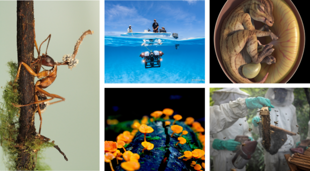
The overall winning image depicts bright orange fruiting bodies growing on deadwood in the Australian rainforest and was taken by Cornelia Sattler from Macquarie University, Australia. The orange pore fungus was first observed in Madagascar but is now found throughout the world. Previous research has reported that invasive species, such as the European rabbit, root rot fungus and feral pigs, threaten 82% of Australian species at risk of extinction. As a result, Australia has particularly strict rules about bringing plants, animals, and organic matter into the country. Cornelia Sattler said: “Despite its innocent and beautiful appearance, the orange pore fungus is an invasive species that displaces other fungi and is spreading throughout the Australian rainforest. It is important to closely monitor this fungus, whose spores are often transported by humans, in order to safeguard the biodiversity of Australia.” Senior Editorial Board Member Arne Traulsen recommended the entry, saying: “Cornelia Sattler’s image allows us to peek into the world of fungi, organisms that are fascinating and yet underappreciated and understudied.”
Research in action: best in category
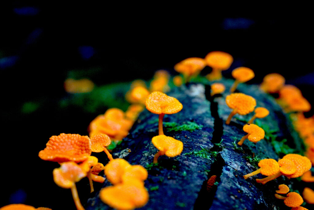
Victor Huertas from James Cook University, Australia took the winning image for the Research in Action category. The photograph depicts the deployment of an underwater remotely operated vehicle at Coral Sea Marine Park, Australia. The device is used to survey oceans at depths that are beyond the reach of divers and has been used to discover new species in reefs and expand the known geographic range of multiple fish species. Senior Editorial Board Member Luke Jacobus said: “This photograph captures the essence of ecological study. It showcases sharp imaging and good storytelling and invites us to be curious about our dynamic world.”
Research in action: runner-up
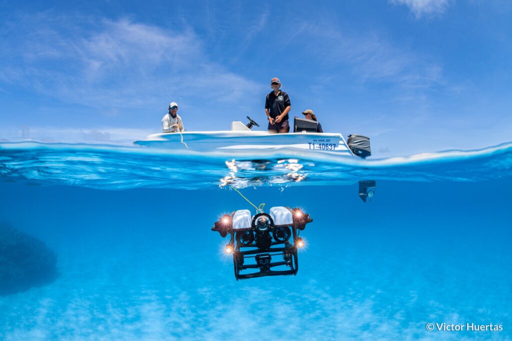
The runner-up for the ‘Research in action’ category captured a moment as researchers conduct a necropsy on a stranded humpback whale that, unfortunately, had passed away. Professor Paul Thompson from the University of Aberdeen in the UK, who submitted the photo taken by James Bunyan from Tracks Ecology explains, “Post whaling recovery of North Atlantic humpback whale populations has led to increases in sightings of this species in UK coastal waters, but this also raises the risk of entanglement in coastal waters. In May 2023, a young humpback whale became stranded in Loch Fleet National Nature Reserve in NE Scotland, drawing the attention of the scientific community. This site has been the focus of long-term photographic studies of harbour seals by the University of Aberdeen, most recently working in collaboration with Tracks Ecology to use UAV (unmanned aerial vehicle) photography to identify and measure individual seals from this population. Colleagues from the University of Glasgow’s Scottish Marine Animal Stranding Scheme conducted a necropsy of this whale, confirming drowning following entanglement was the most likely cause of death. This image was drawn from a series captured by UAV photography, documenting the necropsy and producing a 3-D model of the whale using photogrammetric techniques that were developed to study the size structure of the seal population.” This photo depicts researchers collaborating to further our understanding of the challenges faced by these magnificent creatures.
Protecting our planet: best in category
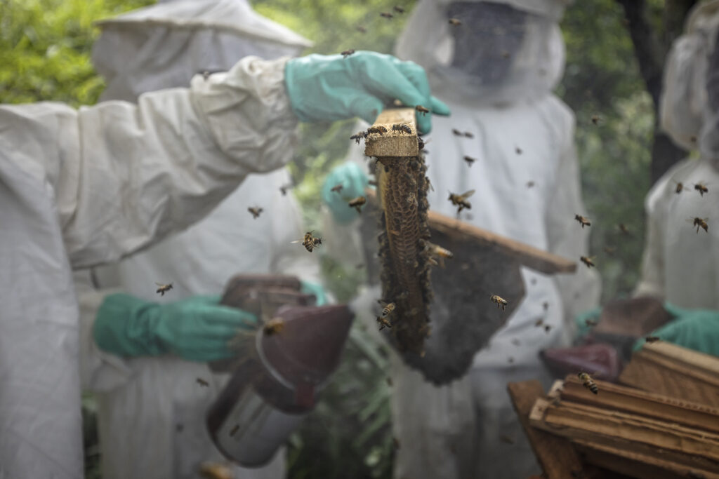
The Protecting our Planet category winner was captured by Roberto García-Roa from the University of Lund, Sweden, and features a sustainable beekeeping project launched by the Chimpanzee Conservation Center in Guinea. The aims to combat deforestation by encouraging locals to cultivate their own honey. A portion of the profits generated by the project go towards chimpanzee conservation activities. Senior Editorial Board Member Josef Settele said: “This photo shows how very different aspects of wildlife conservation can be combined into win-win situation that helps simultaneously protect our planet and empower local communities.”
Protecting our planet: runner-up
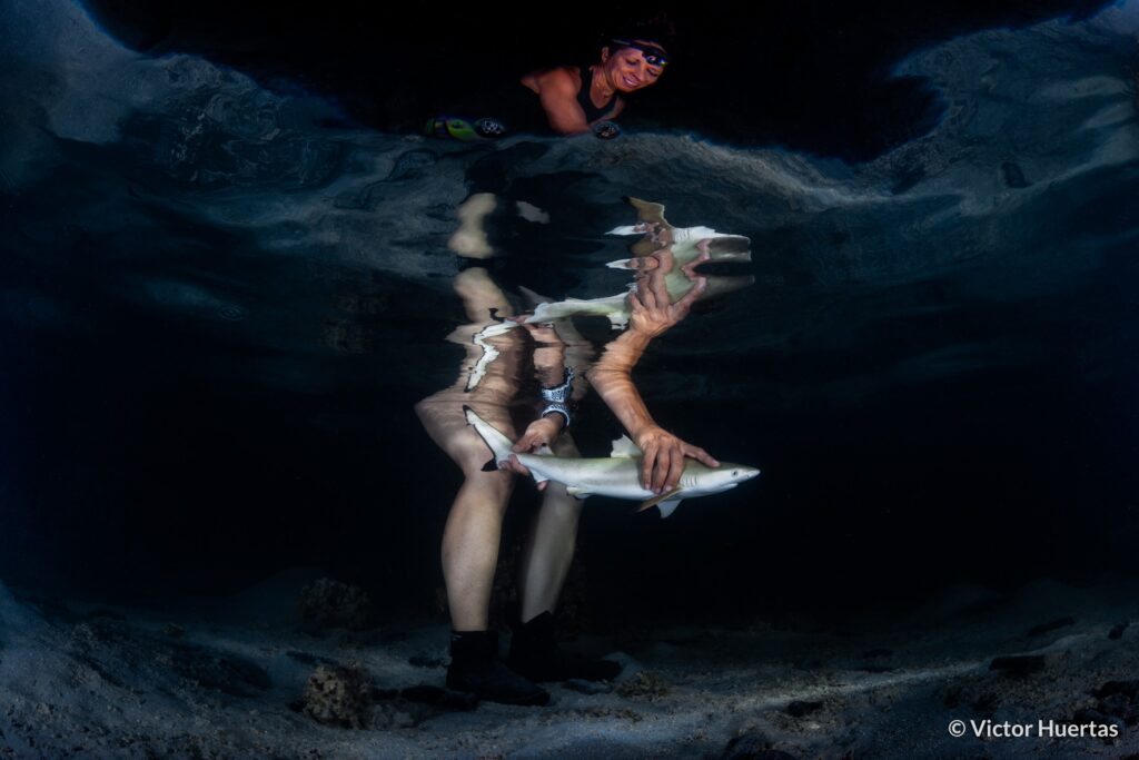
An image captured by Victor Huertas was also selected as the runner-up for the “Protecting our planet” category. The photo captures Professor Jodie Rummer, from James Cook University in Australia, releasing a newborn blacktip reef shark (Carcharhinus melanopterus) in Mo’orea, French Polynesia, after tagging it and collecting biometric data. Victor explains that “Professor Rummer leads Physioshark, a research team headquartered at James Cook University in Australia, that investigates the impact of climate change on the physiological performance of newborn sharks in tropical shark nurseries. These habitats typically occur in shallow waters and are therefore highly exposed to rising temperatures and lower oxygen concentrations. The Physioshark team is untangling the challenges newborn sharks face in such rapidly changing environmental conditions. Professor Rummer and her students have so far been able to show how despite the burden climate change is placing on the physiology of young sharks, these are displaying an exceptional resilience to these changes, giving scientists hope that they will be able to adjust to a warming ocean.”
Plants and fungi: best in category
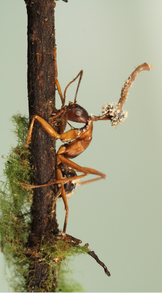
João Araújo from the New York Botanical Garden submitted the winning photo for the ‘Plants and fungi’ category. The image captures a fascinating scene of a fungus parasitizing the fruiting body of a zombie-ant fungus. Zombie-ant fungi possess the remarkable ability to manipulate the behaviour of their insect hosts, compelling them to migrate to a more favourable location for their growth. These incredible organisms infect various Camponotini ants in forests worldwide, from tropical to temperate regions. However, João’s photograph demonstrates that the life of this parasitic fungus is far from simple, depicting the complexity and intricacy of nature. João tells us, “The forests these fungi inhabit are also shared with mycoparasitic fungal lineages that can parasitize, consume and even castrate Ophiocordyceps. Only recently scientists have started to catalogue and describe these still unknown fungi that can kill other fungi.”
Plants and fungi: runner-up
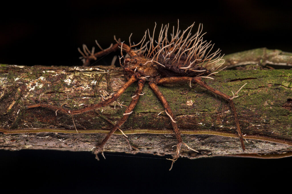
Roberto García-Roa also submitted the runner-up for the ‘Plants and Fungi’ category. This unsettling yet mesmerizing image depicts a spider seemingly defeated by a parasitic fungus. Roberto explains “While it is not uncommon to encounter insects parasitised by “zombie” fungi in the wild, it is a rarity to witness large spiders succumbing to these fungal conquerors. In the jungle, near a stream, lies the remains of a conquest shaped by thousands of years of evolution.” Senior Editorial Board Member Luke Jacobus comments “The image provides an opportunity to expand awareness and understanding of complex and unfamiliar interactions between organisms.”
Paleoecology: best in category
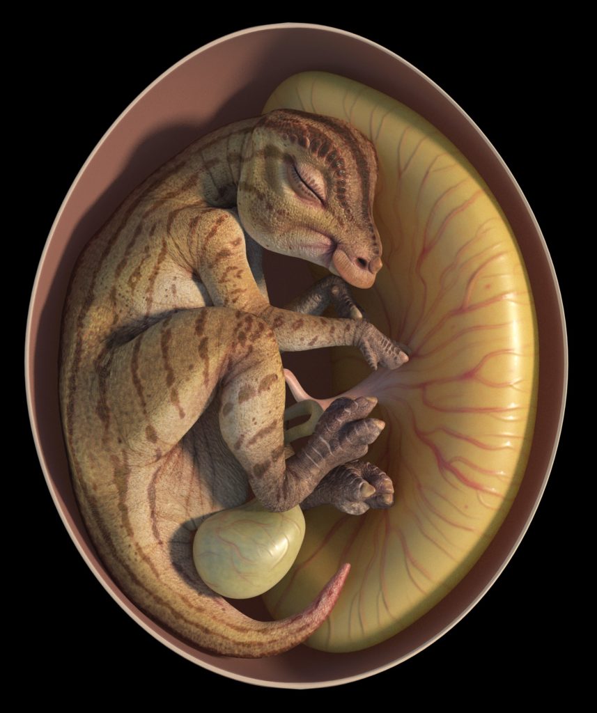
The winning image of the ‘Paleoecology’ category, submitted by Jordan Mallon from the Canadian Museum of Nature, has highlighted the incredible work a group of trans-Pacific palaeontologists completed remotely during the COVID-19 pandemic. Jordan’s submission showcases the team’s collaborative efforts in describing a remarkable discovery: a pair of hadrosauroid dinosaur eggs and embryos from China’s Upper Cretaceous red beds, dating back approximately 72 to 66 million years ago. Jordan comments, “The relatively small size of the eggs, and the unspecialized nature of the dinosaur embryos inside, suggest that the earliest hadrosaurs laid small eggs and hatched altricial young. More derived hadrosaurs eventually laid eggs nearly four times larger by volume and hatched correspondingly larger young. This digital image depicts an example of a ‘primitive’ hadrosaur developing within the safety of its small egg expertly crafted by Wenyu Ren.” This image reminds us of the amazing wealth of information locked within fossils.
Paleoecology: runner-up
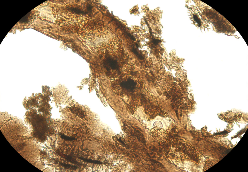
Jasmina Wiemann from the Negaunee Integrative Research Center, Field Museum of Natural History, USA, entered the runner-up for the ‘Paleoecology’ category. Through her microscope, Jasmina provides a view of an extracted diplodocid dinosaur blood vessel surrounded by preserved extracellular matrix containing remnants of cells that are approximately 150 million years old. Jasmina explains, “Once considered paradoxical, the preservation of fragile soft tissues is now known to be the result of the chemical transformation of original proteins, lipids, and sugars occurring during fossilization – allowing such fragile evidence of past life to survive over millions of years!”. The field of molecular paleobiology is important for reconstructing the physiology, relationships and behaviours of long-extinct species.
Many congratulations to all of our winners!
All the figures have been released under a Creative Commons Attribution License (CC BY), so everyone is welcome and encouraged to share them freely, as long as you clearly attribute the image to the author. To learn more about the competition and the winning images, please read our editorial here.

Comments