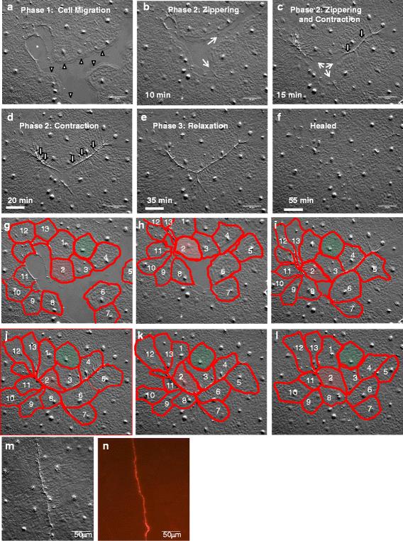
In vivo imaging of epithelial wound healing in the cnidarian Clytia hemisphaerica demonstrates early evolution of purse string and cell crawling closure mechanisms.
The mechanisms of wound healing have always been a highly discussed topic across many fields of science. In their paper, Kamran et al. discuss cell crawling and purse string mechanisms (a supracellular contractile ring that contracts to draw the wound margins inward) in the cnidarian Clytia hemisphaerica, a cousin to the jellyfish. Although these mechanisms are well documented, Kamran et al.’s study presents a fascinating insight into wound healing in vivo.
Using a combination of electron microcopy, staining techniques and various imaging techniques, Kamran et were able to document the wound healing mechanisms in live subjects. As well as developing a wound healing assay in the cnidarian Clytia hemisphaerica, Kamran et al. were also able to establish which wound healing mechanisms occur in this species, discovering that wound healing occurs rapidly and independently of cell proliferation. In addition, they also establish that the implementation of either Lamellipodia-dependent cell crawling or the classical purse string mechanism depends on the integrity of the basement membrane. This fascinating article has shown potential for a Clytia model for wound healing.
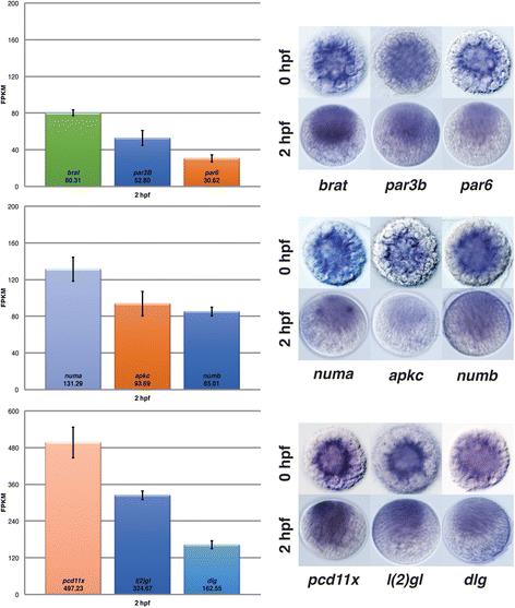
The asymmetric cell division machinery in the spiral-cleaving egg and embryo of the marine annelid Platynereis dumerilii
The importance of asymmetric cell division goes without question. The division of cells within a zygote is imperative to the organization of cellular fates, which, in summary, is essential for the development of organs. Spiral cleavage (the division of cells in a spiral manner), however, only occurs in species such as mollusks and annelids. In their fascinating paper, Nakama et al. sought to analyze the machinery that is involved in spiral-cleaving egg and embryo of the marine annelid Platynereis dumerilii.
During their study, Nakama et al. were able to identify and classify genes that are important to asymmetric cell division in the eggs and early embryos of Platynereis dumerilii by analyzing their stage-specific transcriptome data. They were able to establish that most of the components in the spiral cleavage of marine annelid Platynereis dumerilii are maternal, which suggests that the sprialian egg in this species requires most of the components for asymmetric cell division to ensure rapid cell division. Furthermore, Nakama et al. have effectively carried out a systems-level analysis which has allowed them to identify key regulatory components that are expressed in the Platynereis dumerilii zygote, providing an in-depth insight into the early development of the marine annelid Platynereis dumerilii.
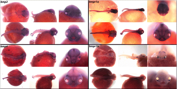
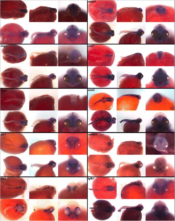
A compendium of developmental gene expression in Lake Malawi cichlid fishes
Despite comparative gene expression being important in developmental and evolutionary research, it seems that spatial expression surveys are restricted to models such as zebrafish and other laboratory models. In this fascinating paper, Bloomquist et al. have produced a compendium of developmental gene expression in a Lake Malawi cichlid fishes, a model that is used to conduct research into the developmental and genetic basis of trait diversity.
In this exciting study, Bloomquist et al. have managed to acquire expression data for many important expression pathways in the Lake Malawi cichlid fishes, including the TGF-β/BMP pathway, FGF pathway and the notch pathway, pathways which are well documented in many laboratory models, including zebrafish, xenopus and the more distantly related chicken. In addition, Bloomquist et al. have also observed stem cell/mitogenic factors, as well as calcium and endocrine signals, at 4 and 6 days post-fertilization, which are rarely seen in whole mount studies such as these.
In summary, Bloomquist et al. have managed to start crafting a compendium which has opened the door for possible future studies into the function of the genes expressed at the early development of Lake Malawi cichlid fishes, which could be applied to many species of Eastern African cichlid fishes.
Comments