This blog was originally posted on the Breast Cancer Now blog.
How did you get involved in cancer research?
Cell biology has always fascinated me, from the early days of secondary school science. Throughout university, I developed a keen interest in cancer research and was lucky enough to do a laboratory placement investigating the role of the immune system in cancer. I went on to study for a PhD, which investigated how proteins known as ‘receptors’ on the surface of cancer cells can encourage growth of a tumour.
I am currently working at the Centre for Cancer Biology in Adelaide, South Australia as a post-doctoral researcher. Still working in breast cancer, I am now investigating how the tumour interacts with surrounding healthy cells, and how this influences growth and progression.
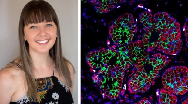
Tell us about your image, Neoplastic Petals
I have been studying how breast cancer develops using a special breed of mouse that develops tumours which closely mimic human breast cancer. My image, ‘Neoplastic Petals’, shows early development of breast cancer in a mouse mammary gland; mammary epithelial cells start rapidly dividing, forming small lesions throughout the gland, which proceed to form small tumours and eventually large, invasive cancers.
As cancer progresses towards the metastatic stage (which is when secondary tumours form), cells become more invasive and mobile. We want to track this process within the cell and to see how different proteins in the cell communicate with each other, with a particular focus on proteins that control cell movement.
How did you create the image?
From the mammary gland tissue of the mice, we took samples which were then preserved and embedded in wax before being finely sliced into thin sections. To make sure our fluorescent dyes targeted the right proteins we used molecules called antibodies – these are a great tool as different antibodies bind specifically to different proteins.
The antibodies were attached to a fluorescent dye which lights up when we view the tissue samples under a special type of microscope, enabling us to clearly see the proteins we’re studying. The different components of the cells were labelled with different fluorescent dyes, allowing us to see the proteins involved in controlling movement (green), the nucleus which contains the cell’s DNA (red) and proteins at the junctions between cells (blue).
How could your research benefit patients and what are your next steps?
We have found that the proteins involved in cell movement (labelled green) are more active during the later stages of cancer, particularly invasive cancer. We think that blocking the activity of this protein, or other molecules that this protein interacts with, could help us to identify new targets which could aid the development of new drugs and improve outcomes for patients.
Going forwards, we want to discover ways to block this protein, which may help us ultimately stop communication between cancer cells and surrounding healthy cells. We think that this protein has an important role in promoting the progression of breast cancer, so understanding how to inactivate it may be key to slowing the rate of cancer growth.
Comments