From mammals to plants
Artificial scaffolds, containing elements with diameters measured at the nanometre scale, have been commonplace in labs studying the behavior of mammalian cells in a tissue-like environment.
Until now, this technology has not come over to the plant science, a reason being the high cost of these scaffolds makes it affordable only to the medi-tech industries. A new method of scaffold production termed ‘shear-spinning’, developed and commericalized by co-author Stoyan Smoukov, now provides mass production of these nanofibre matrices and he has said “with the cost factor now removed, we wanted to test these new scaffolds on plant cells”.
What did we do?
Together with plant scientists from the nearby Sainsbury Laboratory in Cambridge, the team seeded cells from the model plant Arabidopsis thaliana. By first developing the technology for Arabidopsis, we knew we could do much more than observe cells, we could look at various subcellular processes as cells adapted to their new 3D environment.
Once binding and growing between the spaces of the many micro- and nano-fibres their behavior changed dramatically.
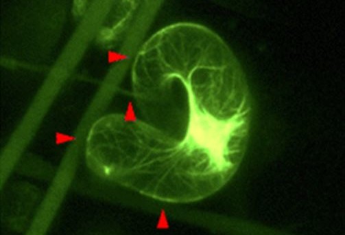
Plant cells can be grown in liquid culture where they adopt regular shapes such as oblong, spherical or kidney-shaped. There was no reason to believe they would be any different in the scaffolds, however, once binding and growing between the spaces of the many micro- and nano-fibres their behavior changed dramatically. The cells were twisting and weaving around their supports in new ways, creating shapes never seen before.
Changing behavior
Clearly culturing the cells in a 3D matrix has changed their behavior. The technique has created an environment that is somewhere between the level of a single cell and that of a single tissue, or organ.
Moreover, these scaffolds are amenable to microscopy. Using fluorescently-labelled reporters, subcellular structures such as the cytoskeleton can be imaged in the growing, shape-transforming plant cell while detailed cell-fibre interactions can be readily probed by scanning electron microscopy.
This is one key application, clearly viewing cell processes in a tissue-like environment. With costs of scaffold production on a downward trend there are also Agri-biotech implications: by targeted differentiation of cells, new biomaterials could be developed.
Comments