Goblet worms, or entoprocts, are rather curious animals. They are sea animals of very small size (0.1-7mm long), that essentially consist of a stalk attached to a rock and a goblet shaped head crowned with a ring of tentacles.
What makes this creature so mysterious?
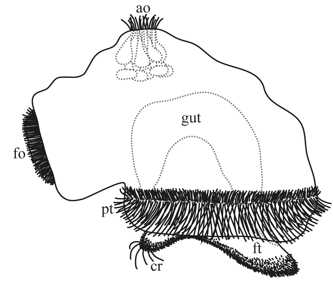
They are unusual in that both their mouth and anus are contained within this crown of tentacles, linked by a U-shaped gut. This is where the name entoproct comes from, literally Greek for ‘anus inside’.
Like most animals that remain stationary as adults, entoproct larvae are themselves very mobile. This is necessary to enable the species to spread and colonise new areas. Interestingly, entoproct sexual reproduction can result in two distinct types of larvae; a swimming type and a rarer ‘creeping’ type that crawls along the sea bed.
Evolutionary origins
Considerable mystery surrounds the evolutionary origins of goblet worms. Some link them to the similar looking bryozoans (or ectoprocts), but others think that molluscs (a group including snails, squid and limpets) are in fact their closest relatives. Resolving such questions is not helped by a distinct lack of fossils and a general lack of understanding of the anatomy of modern day goblet worms.

Attempting to fill this breach is new research, by Julia Merkel and Bernhard Lieb at Johannes Gutenberg University in Germany and Andreas Wanninger of the University of Vienna. Using advanced microscopic staining techniques they attempt to further understand the muscular anatomy of both adults and creeping larvae of the goblet worm Loxosomella murmanica.
The creeping type larvae is thought to be more evolutionary ancient than the swimming type, so the researchers suspected it would give greater insights into common morphology with possible relatives.
The solitary adults of L. murmanica attach themselves to the external surface of the peanut worm Phascolion strombus, which itself lives in shells abandoned by sea snails.
The researchers dredged such shells from the muddy bottom of seas close to Stockholm, detached brooding adult L. murmanica from the peanut worms within and kept the goblet worms in the lab until they released their larvae. Adult and larvae were then stained to detect nucleic acids and actin (one of the main muscle proteins) and examined under the microscope.
Take a look for yourself
Their work, published recently in BMC Evolutionary Biology, has produced some rather remarkable images, which it must be said was one of the main inspirations behind this blog (all images taken from the research paper).
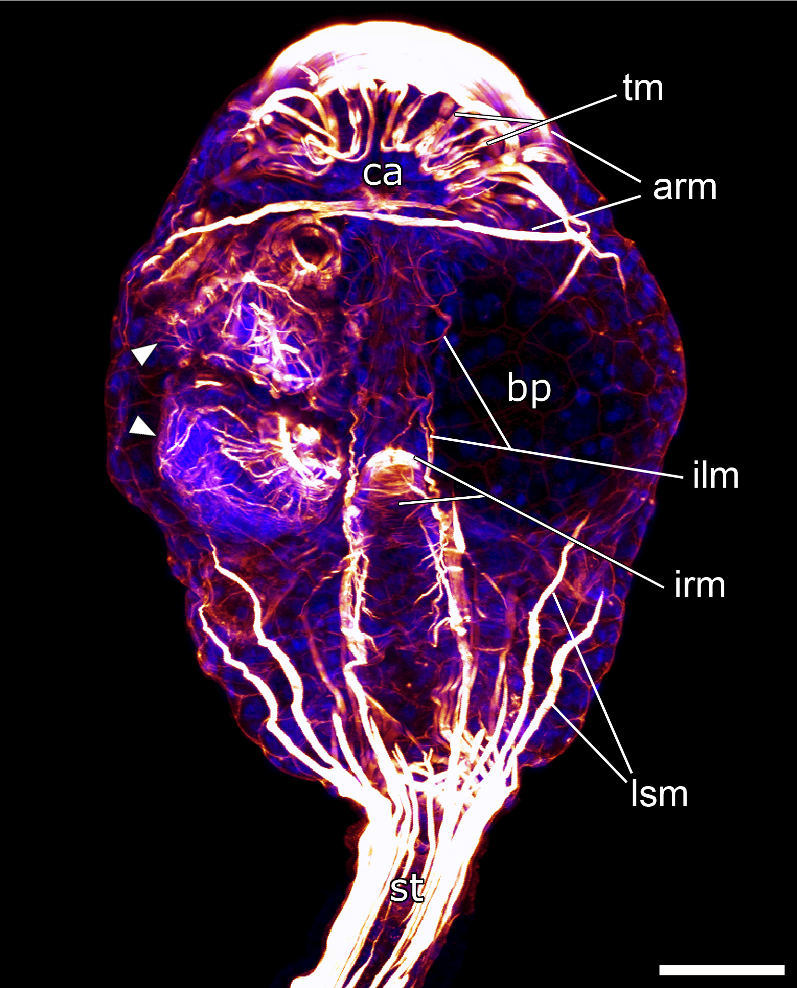

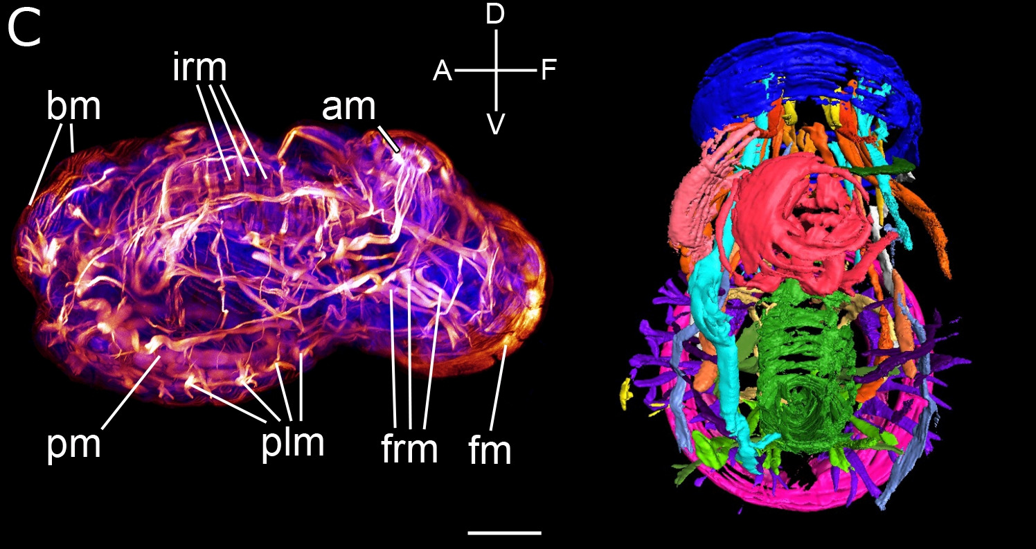
But what do the images show?
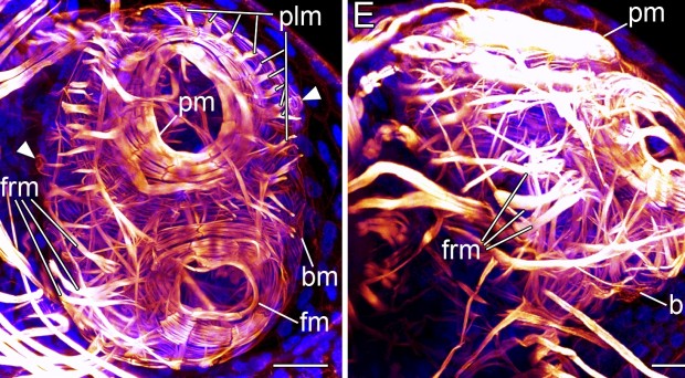
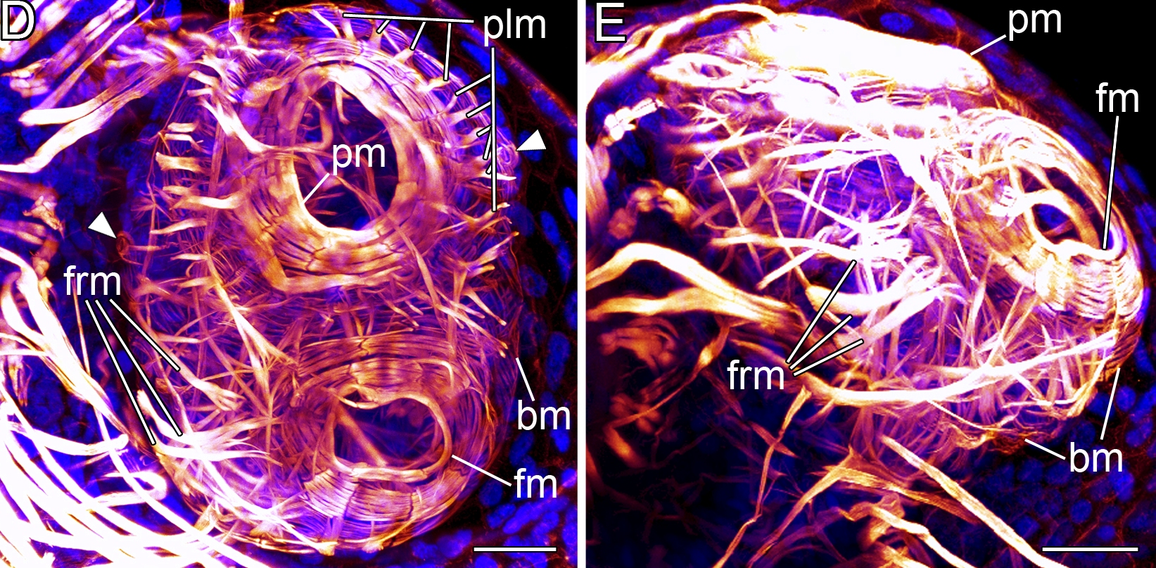
What do the researchers conclude from all of this? Firstly, that the musculature of the creeping larvae of L. Murmanica is surprisingly complex. While some of the muscles resemble those seen in the swimming type larvae, others, like those related to the frontal organ, are unique to the creeping larvae.
Crucially for the question of entoprocts closest relatives, the creeping larvae has very distinct enrolling muscles in the foot structure which are also seen in molluscs, thus supporting the hypothesis that molluscs are the closest relatives of entoprocts.
This research provides an excellent demonstration that modern day imaging techniques allow new insights into the functioning and evolution of previously mysterious species, while still providing images that are beautiful in their own right.
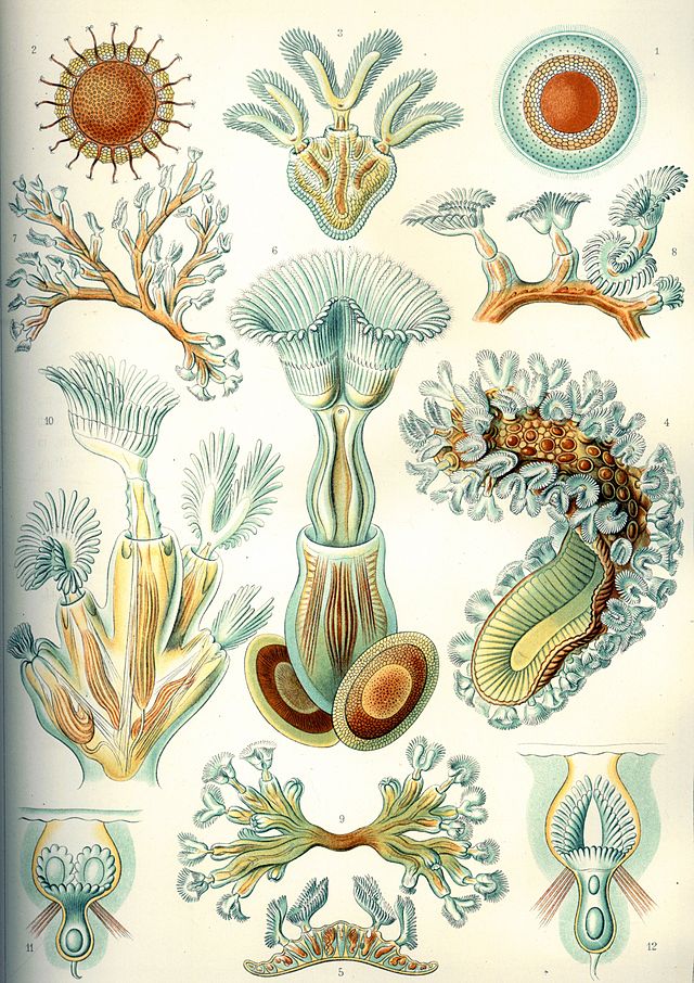
That’s not to say that more traditional ways of visualising the natural world don’t still have their place, so let’s end with some images of the bryozoans (no longer as related to goblet worms as we previously thought) as drawn by legendary biologist Ernst Haeckel over 100 years ago.

Comments