 Commenting on the need for the series, Prof Rodney Hicks (Editor-in-Chief) said “surprisingly, in contrast to the literature regarding the outcomes of imaging, the methods that underpin the generation of an imaging report and how to communicate the findings cogently to referring clinicians remain something of a ‘black box’.
“I hope that you will be as excited as I am to break into data traditionally locked into the brains of experts and only intermittently shared with their own direct trainees or alternatively, at often expensive masterclass presentations”.
In this thematic series, experts across various modalities detail their approach to reporting scans in particular disease settings. In Prof Hicks’ Editorial for the series, he details the motivation for this ‘Master Class Series’ as well as his own approach to reporting.
The series launches with three review articles, two from Editorial Board Members of Cancer Imaging – Prof Stefan Diederich and Prof Harriet Thoeny – and one from former co-Editor-in-Chief Prof Ken Miles.
Use of chest CT in suspected pulmonary complications of oncologic therapies
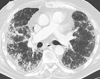
 Prof Diederich’s article entitled ‘Chest CT for suspected pulmonary complications of oncologic therapies: How I Review and Report’ describes imaging strategies and a practical approach to reporting chest CT scan in this setting.
It provides an overview of risk factors, time course and CT morphology of different pulmonary infections, radiation pneumonitis, pulmonary drug toxicity and other pulmonary abnormalities.
Prof Diederich commented “if cancer patients undergoing medical and/or radiation therapy develop pulmonary abnormalities, the differential diagnosis includes progression of malignancy, infection, side effects of therapy and unrelated pathology.
“It is critical to differentiate between these conditions fast and reliably, as they usually require completely different therapeutic approaches and wrong classification may have deleterious consequences.”
Prostate imaging reporting
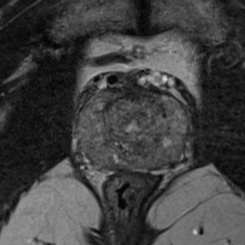
 Prof Thoeny co-wrote a review entitled ‘Prostate MRI based on PI-RADS version 2: How We Review and Report’ with colleague Dr Philipp Steiger.
The aim of their article is to provide an overview of Prostate Imaging Reporting and Data System (PI-RADS™ version 2), how to perform prostate imaging, how to perform image analysis and interpretation and finally, provide insight on how to perform structured reporting.
The authors wrote “In recent years, prostate imaging became more and more popular and is now routinely used in many centers to detect significant prostate cancer. PI-RADS v2 is the newest internationally recognized guidelines for prostate cancer imaging based on multiparametric MRI.
By correctly applying these guidelines, prostate cancer detection will be improved and communication between radiologists and urologists will be facilitated”.
Prognostication of non-small cell lung cancer
 Prof Miles’ review entitled “How to use CT texture analysis for prognostication of non-small cell lung cancer” offers a practical guide to the clinical application of CT texture analysis as a prognostic marker for patients with non-small cell lung cancer.
Prof Miles wrote “too many imaging specialists with an interest in cancer see the application of imaging biomarkers as being primarily relevant to experimental cancer medicine with little bearing on clinical practice.
“By discussing a range of issues such as workflow integration, quality assurance and establishing clinical engagement, this review highlights many of the differences between clinical and research uses of imaging biomarkers in oncology”.
All articles in the series can be read here and further reviews will publish into the series throughout 2016.
Comments