Back in February, we opened up a breast cancer research image competition to anyone affiliated with a research institution or hospital. Entries could be submitted to one of the four categories:
- Immunofluorescence
- Histopathology
- Computer modelling and crystallography
- Other research images
Thanks to everyone who participated! We had so many wonderful entries, which didn’t make it an easy task for the Breast Cancer Now Scientific Advisory Board to pick the winners.
For more background on the winning images, we’ll be posting “Behind the image” posts over the coming weeks, so please be sure to check the On Medicine blog for more details about these amazing images.
Congratulations to our winners!
Histopathology winner – Sarah Boyle, Post-doctoral Research Officer at the Centre for Cancer Biology, Adelaide, Australia
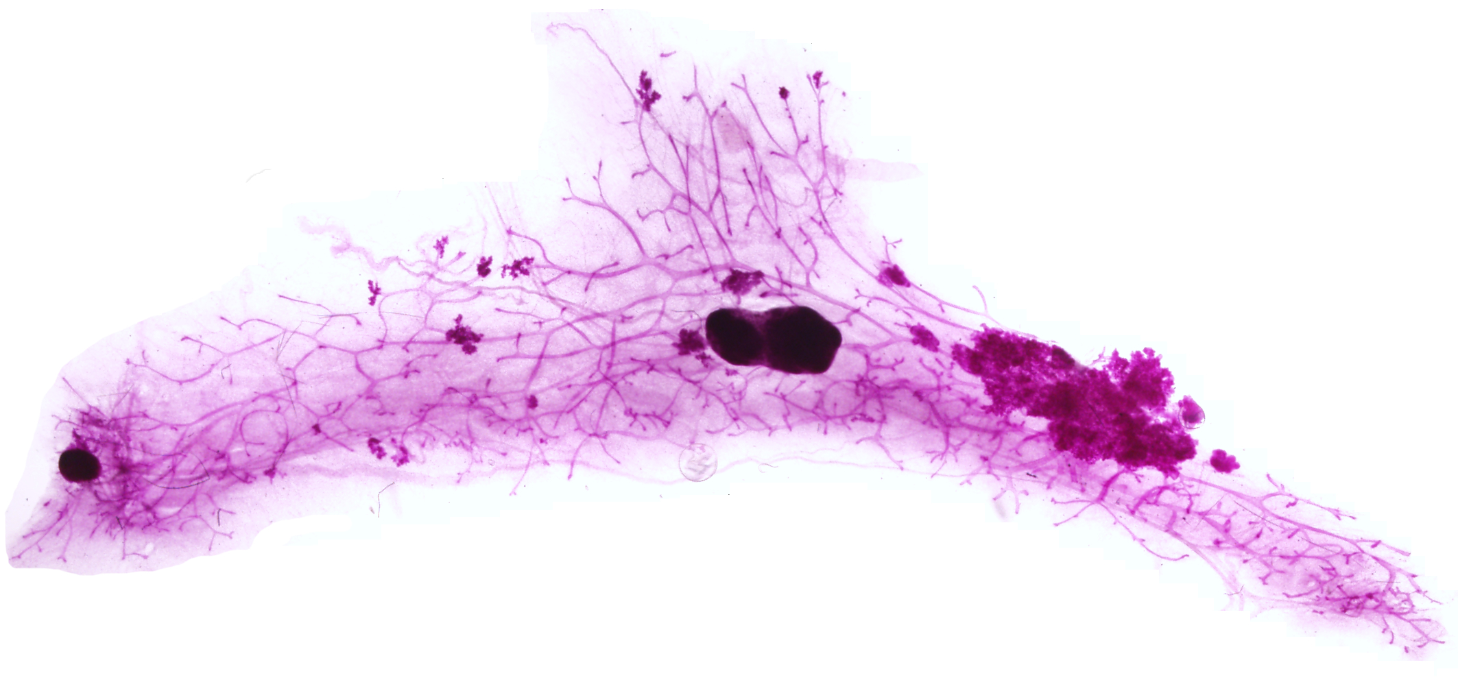
Imaging mouse mammary hyperplasia: This is an image of epithelial hyperplasia (hyper-proliferation of epithelial cells) within a mouse mammary ductal tree. This mouse was genetically engineered to develop mammary tumours, giving us a tool to study development of the disease, closely mimicking human breast cancer. Mammary epithelial cells will spontaneously start to rapidly proliferate, forming lesions like this and those you can see dotted throughout the mammary gland. These eventually go on to form small tumours and then large, invasive cancers that can metastasise around the body. To produce this image, the mouse mammary gland is extracted, spread thinly on a glass slide, fixed, and stained in a dye called carmine alum. The whole mount is then photographed using a dissection microscope.
Computer modelling winner – Edward P Carter, Postdoctoral Research Assistant at Barts Cancer Institute, UK
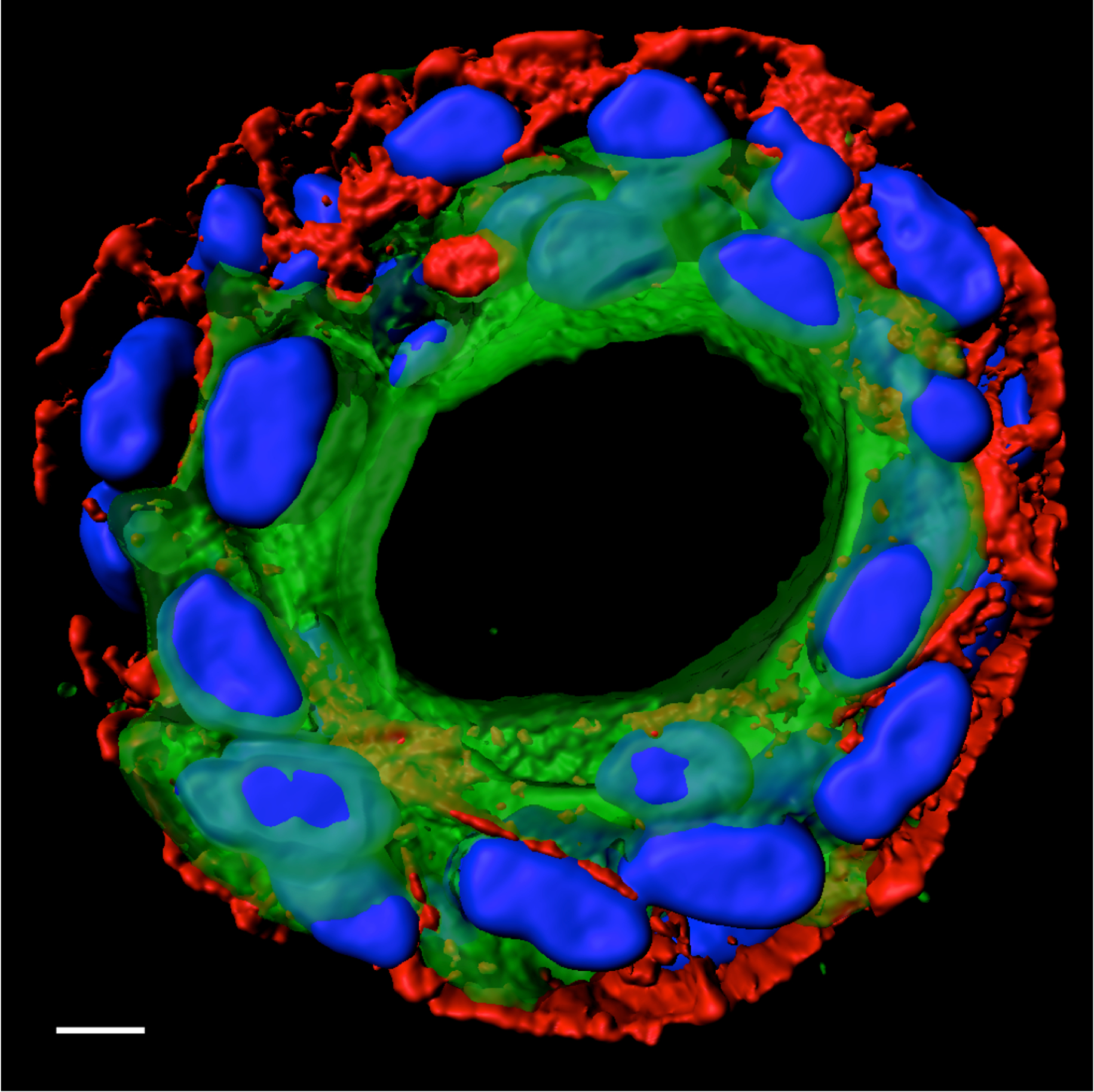
Boob in a tube: 3D model of the human breast duct, developed using using healthy human ductal cells obtained though the Breast Cancer Now Tissue Bank. Cell nuclei are labelled in blue; luminal cells are represented through cytokeratin 8 expression (green) and myoepithelial cells by P-Cadherin (red). Scale bar = 10 μm.
Other winner – James C McConnell, Post Doctoral Research Associate at the University of Manchester, UK
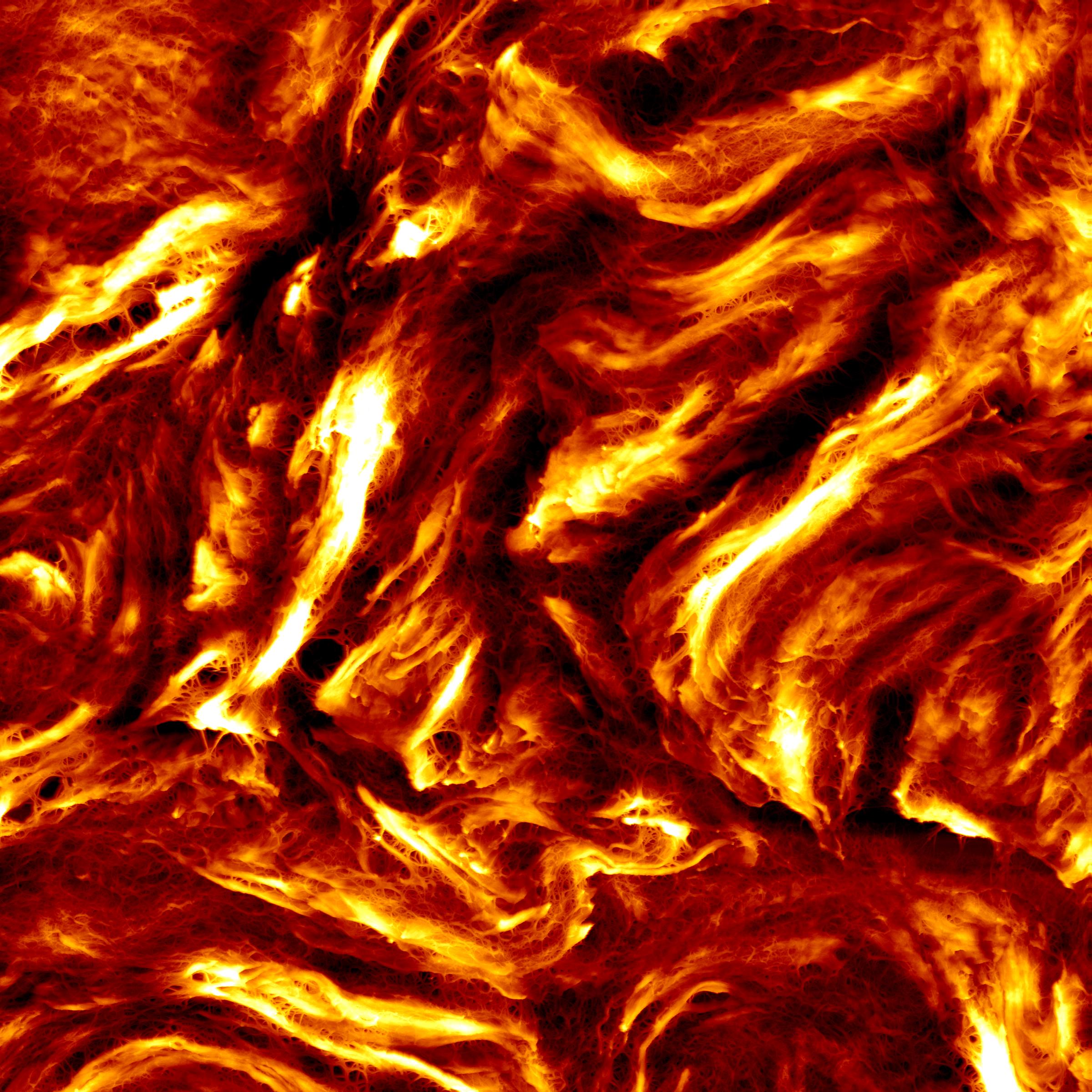
Collagen ultrastructure in high mammographic density breast tissue: This topographical map was generated by atomic force microscopy (AFM) of a sample of breast tissue taken from an individual with high breast density. The map measures 150 × 150 μm, and each line took 12 minutes to collect; the whole map took 16 hours. Large collagen fibril bundles (fibres) can be observed connected to the surrounding tissue by a network of fine fibrils (150–450 nm diameter). Such fibres were not observed in matched low mammographic density breast tissue.
Immunofluorescence and overall winner – Victoria Cookson, Post Doctoral Research Associate at The University of Sheffield, UK
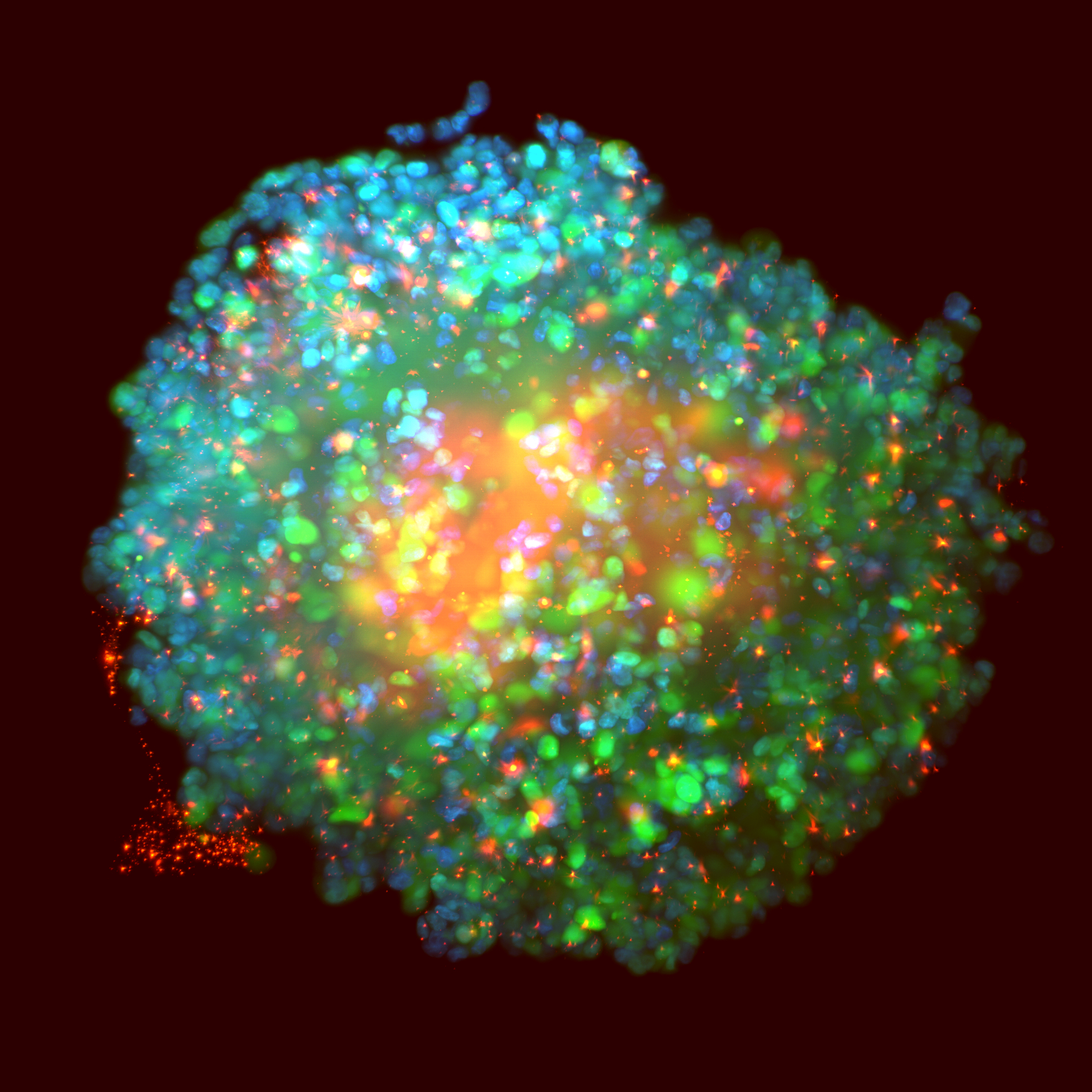
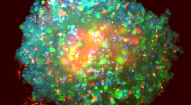
A spheroid generated from a 3D culture of fluorescently labelled MDA-MB-231 breast cancer cells: Fluorescently labelled MDA-MB-231 breast cancer cells (green), with highlighted nuclei (DAPI, blue), transfected with siRNA (red) and imaged using Light Sheet Microscopy. By inhibiting known targets (through either siRNA or small molecule inhibitors) we are investigating molecular pathways involved in breast cancer metastasis.
All images have been released under a Creative Commons Attribution License (CC BY), so everyone is welcome and encouraged to share them freely, while attributing the image author.
Comments