Read the full focus issue here!
Werner Kühlbrandt coined the phrase ‘resolution revolution’ in 2014 highlighting the breakthrough in electron cryo-microscopy (cryo-EM) that allows almost atomic-resolution molecular imaging. Similar advances in high-resolution methods have taken place right across the life sciences, and it certainly hasn’t gone unnoticed, with Nobel Prizes in 2014 for super-resolution fluorescence microscopy, and in 2017 for cryo-EM. This Spotlight draws together some of the recent articles published in the BMC Series, from single cell genomics to light-sheet microscopy.
The transition from bulk sequencing of multiple cells and tissues to sequencing of individual cells has revealed the heterogeneity in the genome, and it’s been used very effectively to map the clonal evolution of cancer. Combining single-cell RNA-seq with bioinformatic approaches has been used to explore resistance to therapy. Single-cell RNA-seq is one of the most popular tools for transcriptomics and this is partly because it can be applied to almost any sample for which a reference genome is available. The software and tools to analyze these data have dramatically increased and the need for careful choice of the pre-processing pipeline is highlighted, as is the need for a method to transform scRNA-seq datasets across platforms.
High-throughput sequencing of trace genetic materials from environmental samples (known as eDNA) is revealing much greater species diversity than previously recorded by either microscopy or historical records. A recent article assessing dinoflagellate assemblages at two different Australian locations showed that sequencing detects rare and less abundant taxa better than traditional methods, and all taxa identified by microscopy were detected by sequencing. eDNA metabarcoding of seawater samples was effective in detecting shark and ray species, and compared with natural history collections and previous survey reports reveal a new degree of species diversity. Further examples of the progress in the application of eDNA (and eRNA) in ecology can be seen in the collection in BMC Ecology and Evolution.
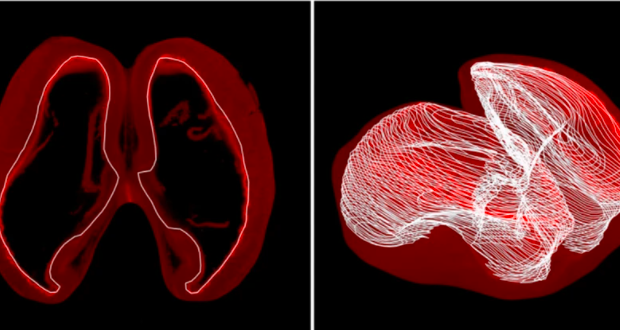
Microscopy remains important, especially at a cellular level, particularly when combined with other digital or technological innovations. Visualization of tumor tissues in three dimensions through virtual reality offers a new approach to teaching and studying pathology. And light-sheet microscopy, a fluorescence microscopy technique with good optical sectioning, was used to characterize embryonic mouse brains in a potential model of autism spectrum disorder. Using 3D printed micro plate inserts has allowed long-term high resolution imaging of whole live brain organoids.
Although cryo-EM has stolen the limelight for advances in molecular structure, other methods continue to improve and particularly to be used to study molecular interactions in live organisms. For example, atomic force microscopy has revealed the mucosal surface properties of a salmon fry without the need to remove the mucosal surface, and NanoBRET (bioluminescence resonance energy transfer) has been applied to the nematode C. elegans in vivo for the first time to detect protein-protein interactions.
We hope you enjoy these highlights and we encourage submissions across the breadth of genomics, ecology, cellular biology and molecular biology as well as plant biology, microbiology, biotechnology and biomedical engineering.

Comments