
Earlier this year we proudly announced that BioMed Central is becoming BMC. Firmly believing that our research communities share our enthusiasm for innovation, science and progress we launched our first ever “Research in progress” photography competition. We asked you to send us inspiring images reflecting curiosity, integrity and innovation across four categories: people at work, close-ups of equipment, plants and animals and microscopy, and you certainly didn’t disappoint.
So without further ado, here is our winning image, the runner up and a selection of images that caught the eyes of our judging panel of editors and designers.
Winning image
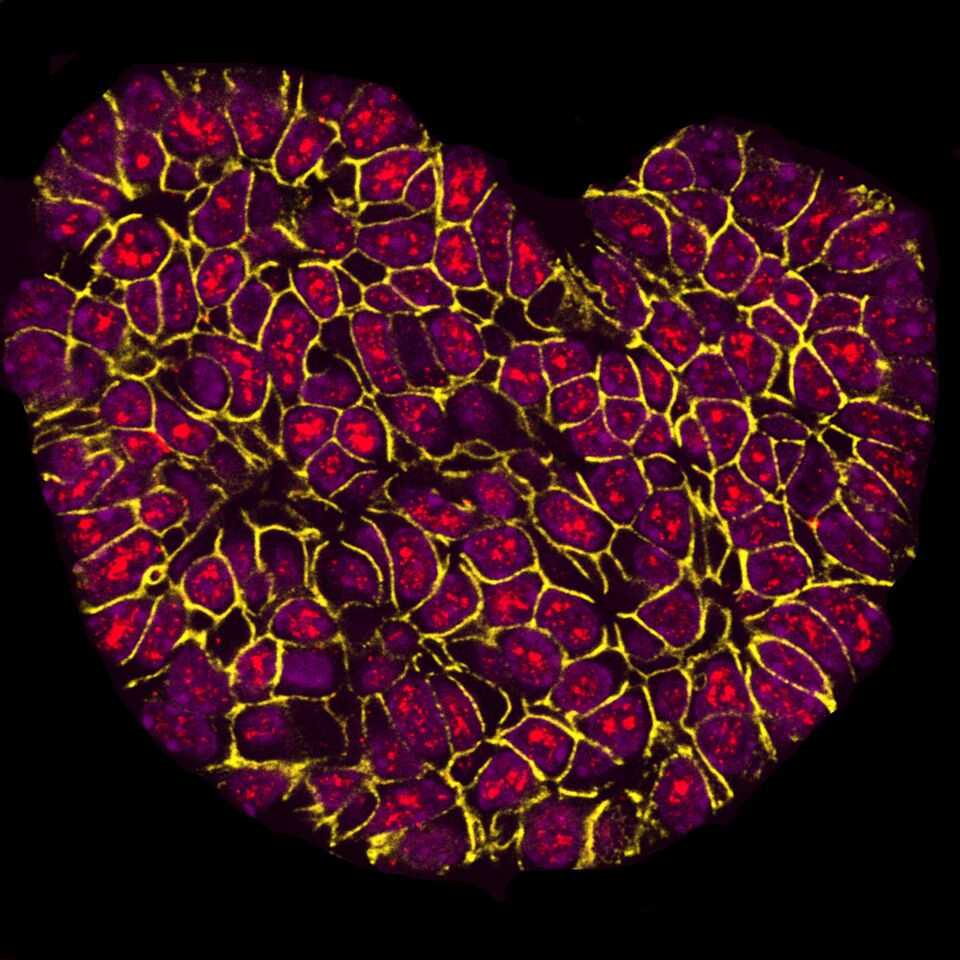
This photo shows a fluorescently labeled mouse mammary tumor produced by scientists studying the progression of breast cancer. The red color labels the active form of a protein, as cancer develops the levels of this protein may increase.
Speaking to us about her entry, Sarah said: “I’m delighted that the image I submitted has been selected as the winner. I took it as part of my research into breast cancer and for me it really shows how processes that we researchers use almost on a daily basis – such as fluorescent labeling and microscopy – can reveal stunning shapes and colors in things like human cells.”
Runner up
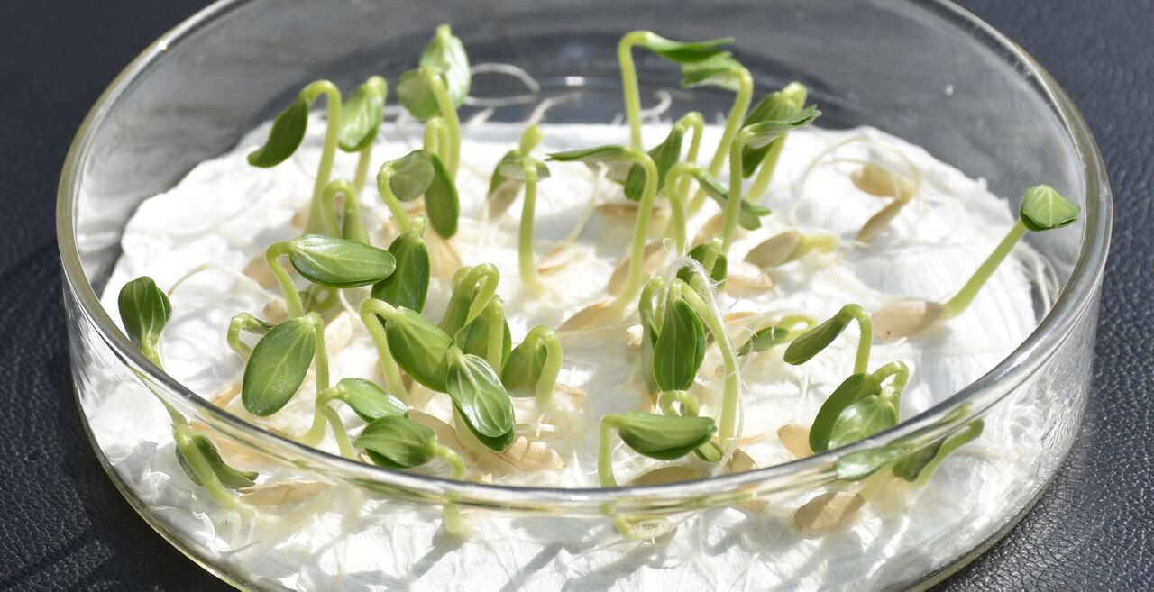
This photo shows cucumber seeds growing in a petri dish. This experiment tests how well seeds germinate and how fast they are likely to grow and establish crops, which are important factors in cucumber breeding.
Selected images
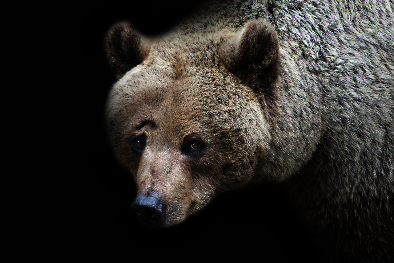
“While traveling Sweden, I visited a small zoo in the mountains; this is where I came across this magnificent bear and her cubs. She had such an intense stare with curiosity and fear in her eyes, she was beautiful. I hope one day she will know what it is like to be free.”
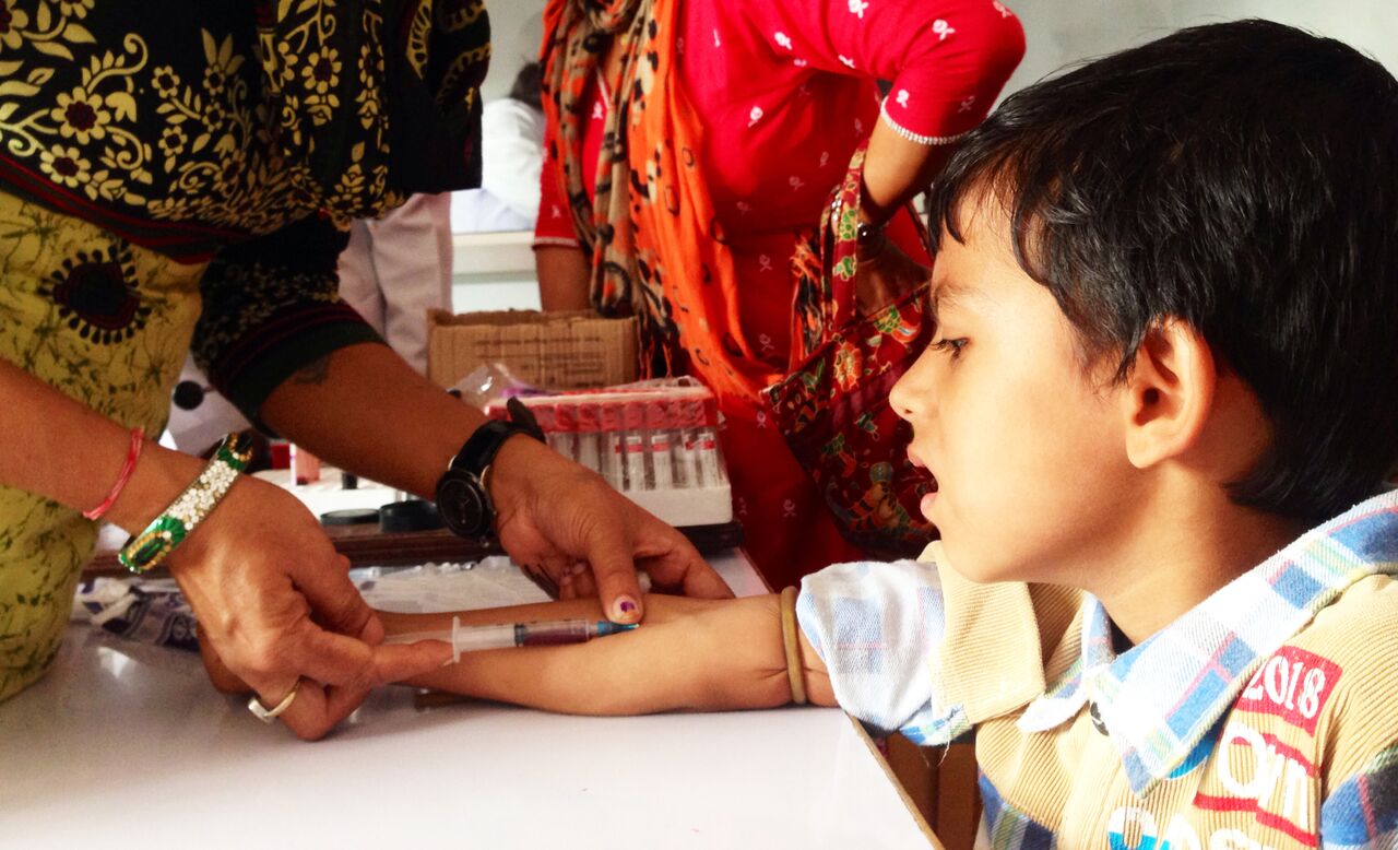
A dengue-suspected child has their blood sampled in the Microbiology/Pathology lab of Ratnanagar Hospital, Nepal. Taken in 2016 amidst epidemicity.
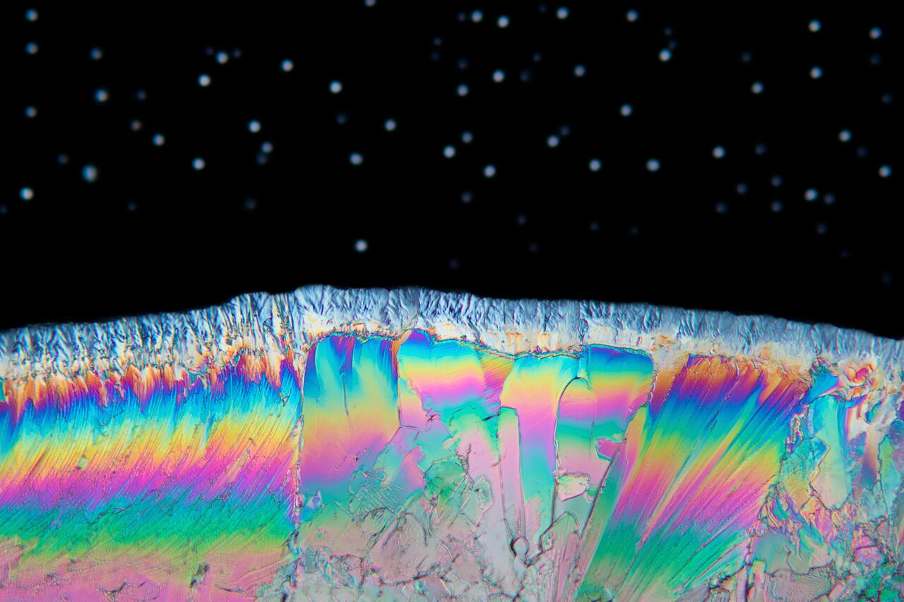
“This image is actually a sliver of a thin film of acetaminophen, commonly known as Paracetamol, imaged to a microscope configured for polarised light. The stars in the background are really just out water droplets that lie in a different focal plane.”
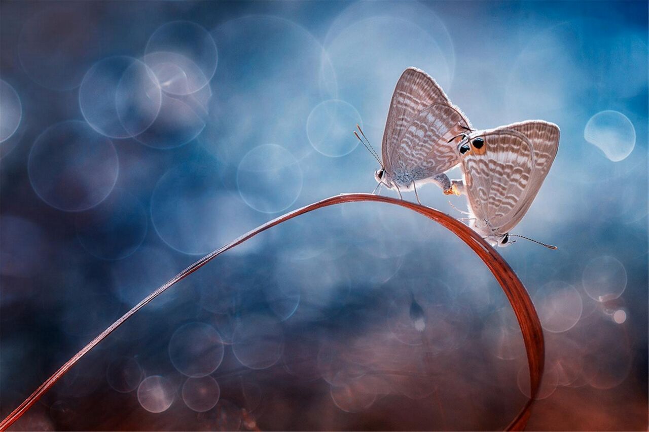
Butterflies, energy-saving.
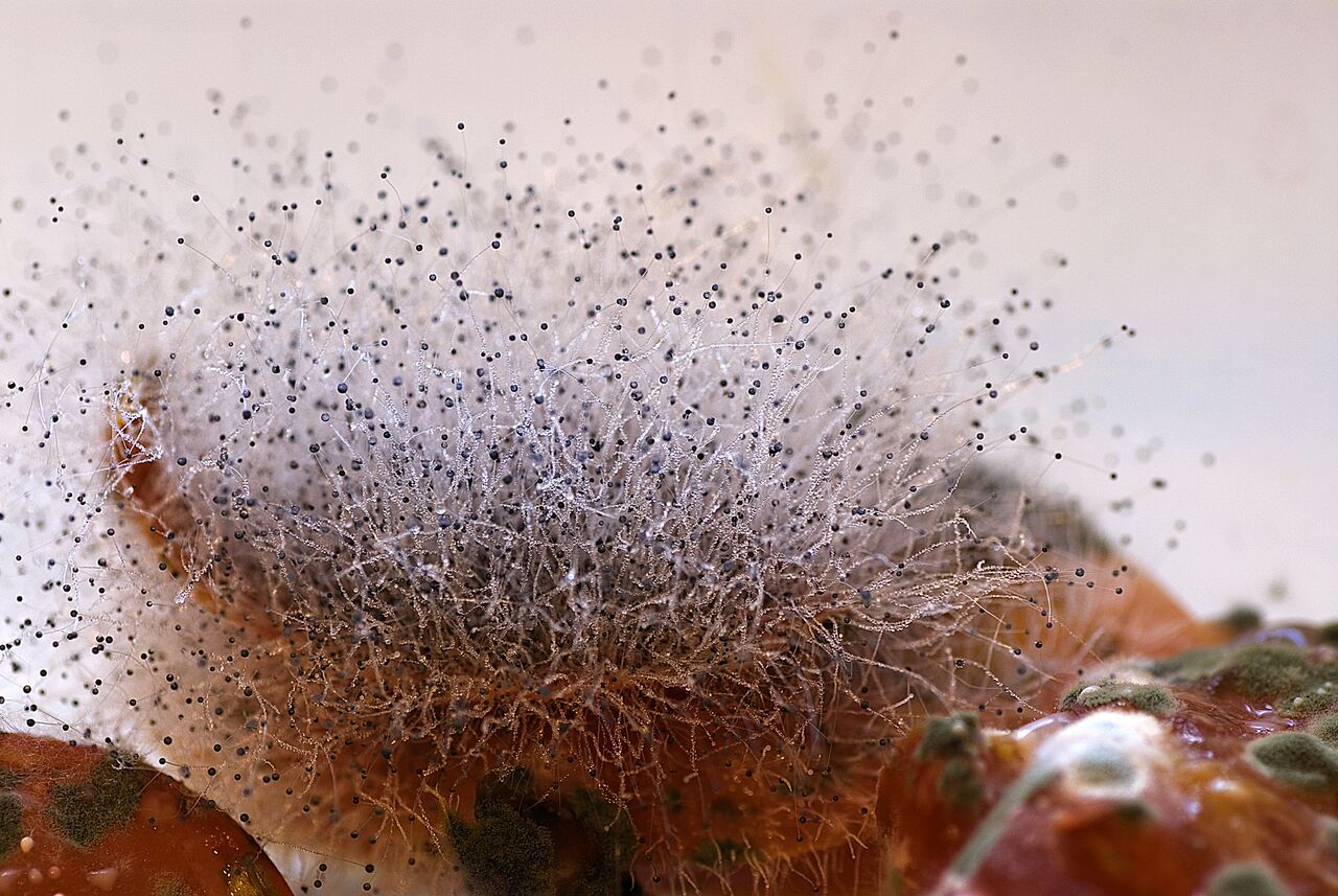
“It is hard to stay away from moulds. They follow you from the petri dishes in your lab to the shelves of your fridge at home. It was here that I saw this one on the reddish background of a decaying tomato. I thought that it was worth of a macro shot before deciding if it deserved a role in research too.”
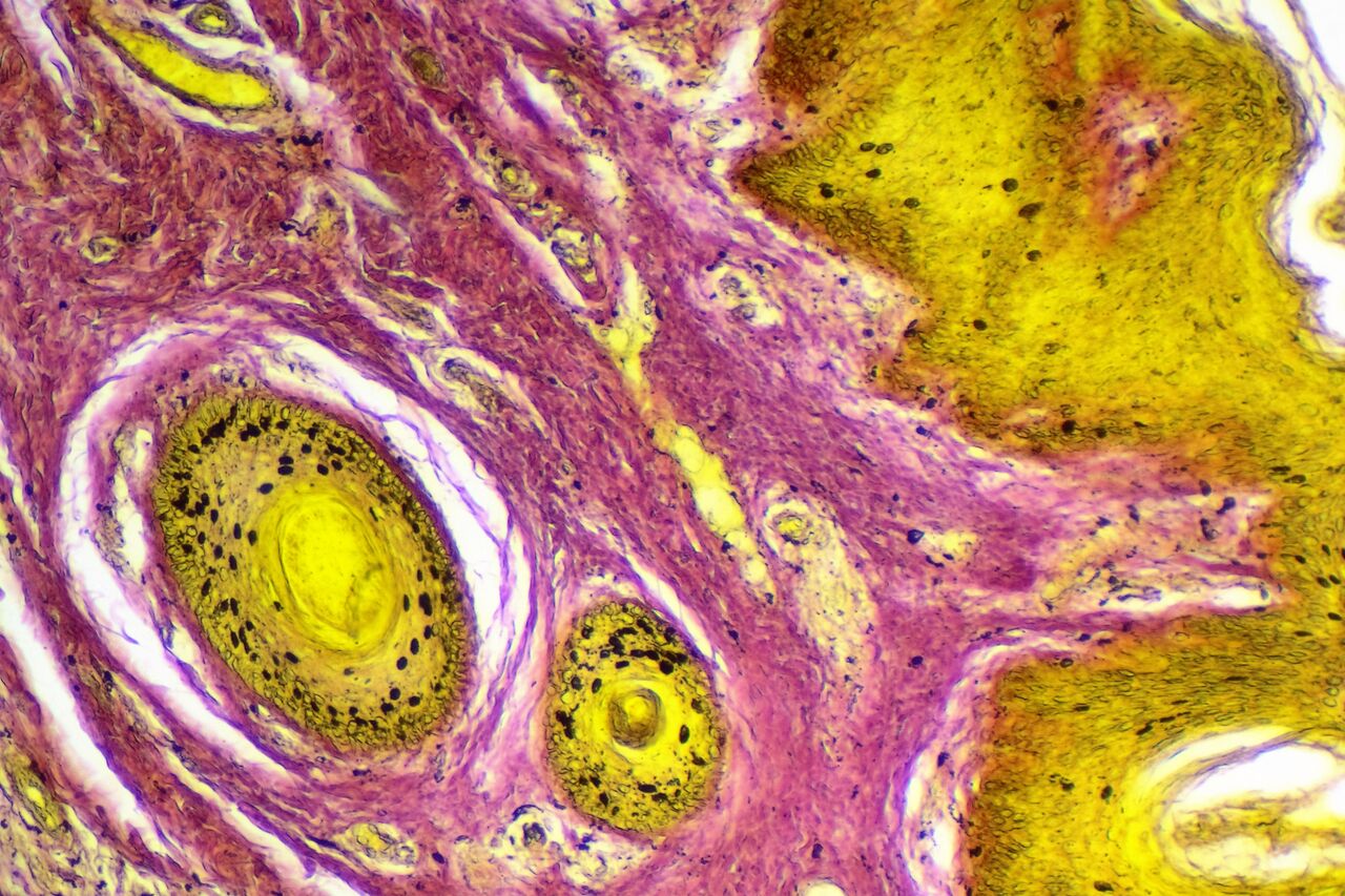
“I have discovered a new stain for detection the mitotic activity in cancers, all the colors in this image are made by my stain. The nucleus with mitotic activity had been stained by black color, the cytoplasm with yellow color and the collagen fibers stained with pink color. This stain colored the epithelia yellow and stroma pink. You can also see the nucleus with mitotic activity colored by black color in both epithelial and connective tissue.”
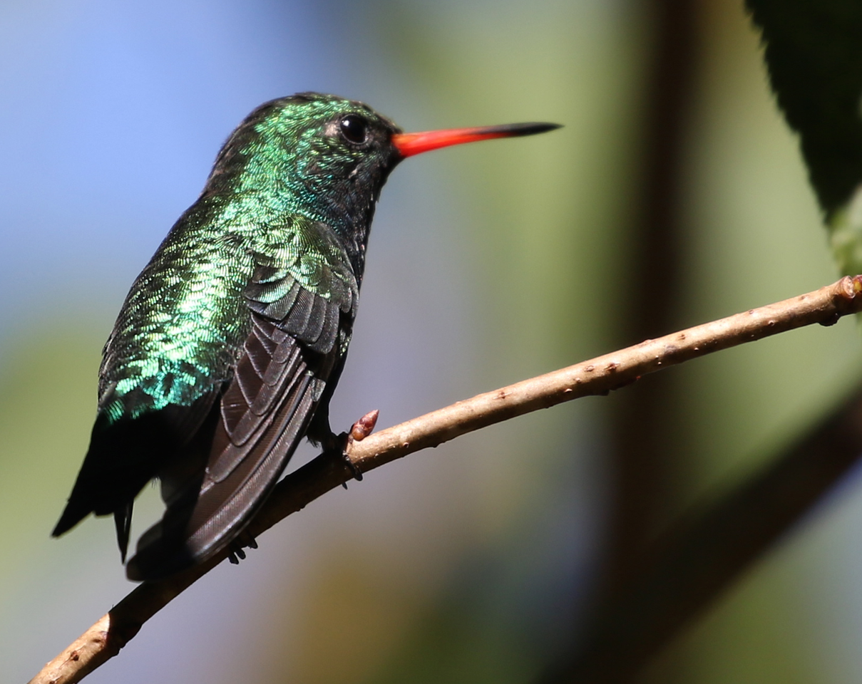
“This photo is part of research entitled ‘e-Nature: validation of images of nature for clinical use in hospital setting.’ It is a methodological study to validate the images, as well as to know the valence and arousal ratings of each type of image. The study will be complemented by a randomized clinical trial on the impact of nature imaging on states of mood and adverse events of chemotherapy in cancer patients.”
We want to say a huge congratulations to the two winners and a massive thank you to all those who entered. Our judges were astounded by both the volume and quality of the submissions.
All images have been released under a Creative Commons Attribution 4.0 License, so everyone is welcome and encouraged to share them freely, while attributing the image author.
Thanks for your cooperation and we will interested to share with you next time.good luck for all winners
Congratulations to all the winners!
We had received an email from BMC informing us that our entry ‘Research by the brook’ has been chosen in the Special Selection category. However, no further information has been provided regarding posting of the photo on the website etc. We kindly request further information as we would like to share it with colleagues and partners. Thank you.