You may have experienced the disconcerting and uncomfortable sensation of an electric shock or even had an electroencephalogram (EEG) test, which monitors the electrical activity of the brain. The interaction between electricity and humans has become relatively commonplace, what’s less well known is that electrical signals also exist within humans. Bioelectricity, electricity inside living organisms, was discovered over 200 years ago with the German scientist Luigi Galvani providing the first evidence for “animal electricity” In 1794.
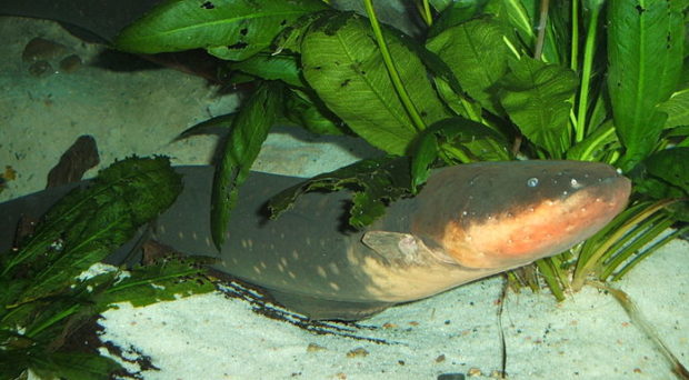
It has since emerged that bioelectricity occurs ubiquitously in the animal and plant kingdom. There are at least 500 species of electric fish, the most famous being the electric eel. Adults can weigh up to 20 kg and can produce a shock of 500 volts, more than enough to kill a human.
How does bioelectricity affect cells?
We now know that living systems exhibit a remarkable ability to utilize electric potentials in cell self-assembly, remodeling of complex shapes, wound healing and tissue regeneration. However the way in which cellular electric potential networks construct and repair tissues remains an open question.
Studies in the last decade have provided convincing evidence that there is a role for electric fields in wound healing.
Electric potentials direct cell migration. This can be seen in nearly all mammalian cells when placed in an external electric field (EF), a phenomenon termed electrotaxis or galvanotaxis.
Electrotaxis is the directional movement of cells along an electric field. Naturally occurring electric currents in the wound EFs have remained poorly understood or largely ignored; but we can use a vibrating probe, glass and platinum microelectrodes or Bio-Electric Imager® to detect and visualize electric potential at the skin surface.
Studies in the last decade have provided convincing evidence that there is a role for EFs in wound healing. It is intriguing that an electric field is able to reverse or over-ride cell migration in a scratch wound model.
This model consists of a single layer of cells with a scratch wound; normally cells on the sides migrate into the empty space created by the scratch. However, application of an electric field can override this process and cause the cells to migrate in the opposite direction.
Unexpectedly, externally added electric fields appears to be a more powerful and dominant cue, overriding other guidance cues in a given cell movement assay. Detailed molecular mechanisms that involved in electrically guided cell migration include field guided positioning of cellular organelles, molecules termed lipid rafts, charged polyamines and ion channels. All of which are responsible for but not exclusively involved in guiding cell migration, by providing spatial sensing of electric field and voltage gradients.
How are we using bioelectricity for healing?
Utilizing bioelectricity provides an unprecedented opportunity for the development of therapeutic strategies to augment the wound healing process and tissue regeneration. Electrical stimulation acts on all levels of tissues and organs, and at each stage of wound healing.
There is compelling evidence of electrical stimulation activating wound healing, and clinical trial data fueling the development of novel devices such as wound dressing product Procellera™, which is electrically inactive in dry conditions and activate when its batteries are moistened by wound exudate or by saline. Other non-invasive, electrical stimulation devices are in development, enabling the transfer of a small current to surface of wound.
In this review, we analyzed the clinical trial data and found encouraging results – chiefly, a significant improvement in chronic and non-healing wounds following electrical stimulation. The beneficial effects of electrical stimulation on wound healing can no longer be ignored.
The mechanism of electrically stimulated cell migration and its unique overriding effects over other migration cues is unprecedented. In this study, we highlighted our collective effort in the search for cellular mechanisms of galvanotaxis. This included using methods to visualize the activation and polarization of the PI3K kinase/Akt pathway, an important signaling pathway in regulating the cell cycle, in a neutrophil-like cell in an electric field.
Our most recent effort in the search for cellular targets screened a library of over 300 genes encoding ion channels, pumps and transporters using a siRNA knockdown strategy on a human corneal epithelial cell line. Over the past 150 years, we might have only scratched the surface.
Endogenous electric fields were detected at human skin wounds over a century ago; however its role remains largely unknown. Bioelectricity is an exciting emerging discipline. “We can increase the natural electrical signals using a variety of chemicals we’d apply to wounds, and by doing that we can get faster healing. What amazes me is that this has been relatively neglected for such a long time,” said. Professor Colin McCaig – Head of School of Medical Sciences at the University of Aberdeen, Scotland.
Luigi Galvani (1737-1798) was in Italian (not German ! ) anatomist, physiologist and physicist. The experiment with the response of frog muscles to electric stimuli was made by him in 1771.
Hii