Like many things in life, biology experiments require balance. The researcher must balance the ease of the experiment with the relevance of the result. As a researcher becomes more reductionist in their experimental design, the experiment becomes more feasible.
For example, a nephrologist could design a study of kidney function within a model organism, in isolated kidneys, or in a kidney cell line grown in a petri dish. In fact, she can even study certain reactions in purified proteins in vitro.
With each of these reductions, however, as ease increases (smaller sizes, fewer complicating factors, less variability), the physiological impact of the result becomes more convoluted. Do cultured kidney cells behave exactly the same as kidney cells within a functioning kidney? Surely not.
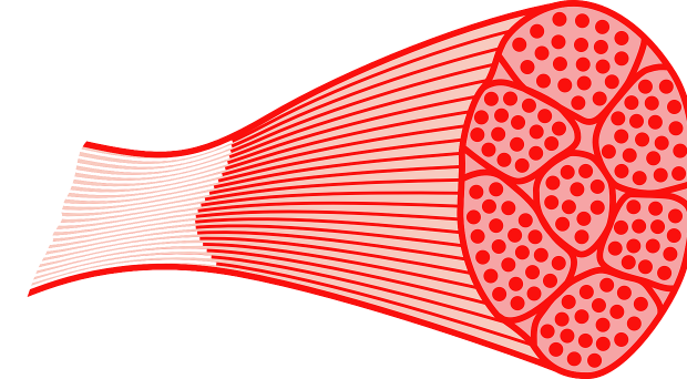
A recent Skeletal Muscle manuscript by Olsson et al. confronts the issue of ease versus relevance in muscle fibers. The functional unit of human skeletal muscle is the muscle fiber. Researchers can learn valuable lessons about muscle biology and diseases by studying muscle fibers isolated from patients with musculoskeletal diseases, as well as healthy controls.
Differentiated myotubes
Intact muscle fibers can be dissected from humans, but this technique is difficult, often resulting in a very small numbers of viable fibers. While just a few viable fibers may be usable for certain techniques (imaging or patch clamping studies), others (such as biochemistry) require larger numbers of cells.
Therefore, researchers will isolate muscle precursor cells from biopsies, and then differentiate these into myotubes in culture dishes. These myotubes serve as a model for human adult skeletal muscle. Myotubes appear very similar to isolated muscle fibers – both are elongated, multinucleate and contract upon electrical stimulation. For these reasons, differentiated myotubes have become a commonly used tool for studying many of the molecular and physiological properties of muscle.
The importance of calcium
Olsson et al. chose to directly compare the calcium transients and subsequent contractions observed in myotubes to those observed in muscle fibers.
Regulation of intracellular calcium is crucial to skeletal muscle function. For one, the global wave of calcium that floods the muscle cell in response to membrane depolarization (aka the ‘calcium transient’) acts to couple surface membrane depolarization to muscle contraction.
And the intrinsic speed of the calcium transient is in turn modulated by the presence and localization of calcium-handling proteins. Olsson et al. chose to directly compare the calcium transients and subsequent contractions observed in myotubes to those observed in muscle fibers.
They found marked differences between the two. Upon electrical stimulation, nearly all of the intact muscle fibers showed transient increases in calcium and force-producing contractions whereas only about 50% of myotubes showed similar transient increases in calcium upon stimulation.
Further, the kinetics of these increases were altered, and they were not always accompanied by myotube contractions. The reason for this triggering misfire becomes clear as the authors go on to show that myotubes lack the arrangements of actin and myosin filaments necessary for contraction to occur. Additionally, a number of important calcium-handling proteins are downregulated or mislocalized in myotubes, compared to muscle fibers.
What can researchers do?
Is there anything a researcher can do to encourage a myotube to be more similar to a fiber? The authors of the current study do not address this experimentally, but previous work from Tanaka et al. show that human muscle cells that were innervated by being co-cultured with rat spinal cord showed continuous contraction and extensive cross-striations similar to intact muscle fibers. Innervation is likely the key to making a myotube more like a muscle fiber.
Overall, these results provide a cautionary tale for muscle researchers.
For researchers interested in the functional studies of human muscle fibers, innovation will be required. Olsson et al. demonstrate reasonable success in isolating fibers from intercostal muscle biopsies obtained during thoracotomies. The intercostal muscles are particularly amenable to fiber isolation due to the relatively short lengths of the fibers (0.5 – 1 cm). Biopsies from muscles containing longer fibers (such as limb muscles, which can be up to 30 cm) will likely prove difficult to adapt to this technique.
Overall, these results provide a cautionary tale for muscle researchers: myotubes are not an adequate model of adult muscle fibers when studying the details of calcium dependent processes. Future studies could likely show marked differences between myotubes and muscle fibers in additional cellular processes, as well.
Jennifer Levy
Latest posts by Jennifer Levy (see all)
- Model behavior? Myotubes and the quest for an ex vivo model of muscle - 6th October 2015
Comments